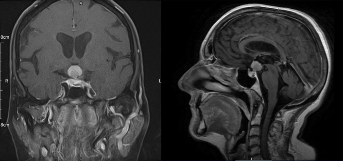
Figure 2.
Head MRI with contrast (coronal T1 FS and sagittal T1 FLAIR) after 6 weeks demonstrating a significant increase in the size of the sellar-suprasellar mass now measuring 22×14×15 mm, with the largest portion lying in the hypothalamus, and evidence of impingement affecting both optic nerves and the optic chiasm

