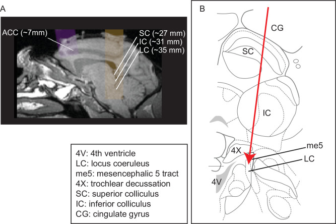
Figure 1. Recording site locations.
(A) Approximately sagittal MRI section showing targeted recording locations in the anterior cingulate cortex (ACC) (areas 32, 24b, and 24c) and locus coeruleus (LC) for monkey Ci (right), with the SC and IC shown for reference. For recording locations in monkeys, Oz, Sp, and Ci (left hemisphere), see Kalwani et al., 2014; Joshi et al., 2016. (B) Schematic of a coronal section showing structures typically encountered along electrode tracts to LC (adapted from Paxinos et al., 2008; Plate 90, Interaural 0.3; bregma 21.60).

