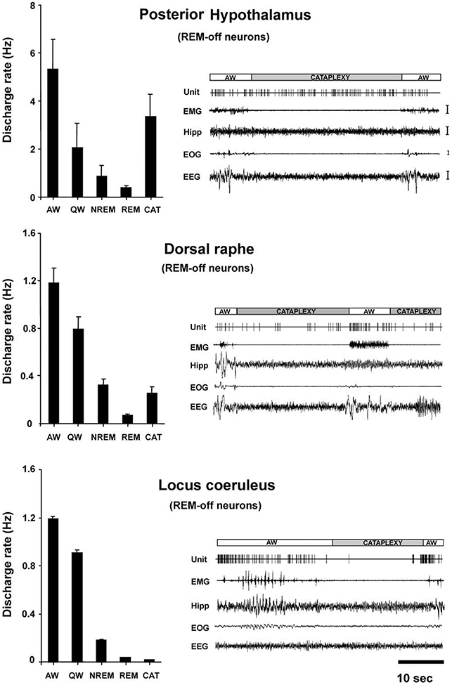
Figure 4. Comparison of Mean Discharge Rates and Cataplexy-Related Activity of REM-Off Cells Recorded from Three Brain Regions.

Posterior hypothalamic (histaminergic) neurons remained active, whereas dorsal raphe (serotonergic) neurons reduced discharge (Wu et al., 2004), and locus coeruleus (noradrenergic) neurons ceased discharge (Wu et al., 1999) during cataplexy. All of these cell types were active in waking, reduced discharge in NREM sleep, and were silent or nearly silent in REM sleep. Calibration: EMG, 100 μV; Hipp, 50 μV; EOG, 50 μV; EEG, 50 μV. Abbreviations as in Figure 2 and 3 legends.
