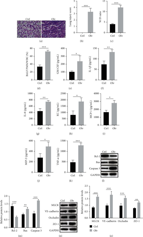
Figure 1.

OLV induces inflammatory activation and lung permeability changes. (a, b) H&E staining detected the pathology of lung tissues in OLV and control rats. (c) The lung W/D ratio between OLV and control rats was calculated according to the formula [weight (wet) − weight (dry)]/weight (dry). (d) The PMN/WBC ratio of BALF in OLV and control rats was calculated. (e–k) ELISA performed to examine the levels of inflammation mediators (GM-CSF, IL-1β, IL-8, KC, MCP-1, MIP-2, and TNF-α) between the OLV group and control rats. (l–o) Western blot detected the protein levels of Bcl-2, Bax, caspase 3, MLCK, VE-cadherin, occludin, and ZO-1 in OLV and control rats. ∗P < 0.05, ∗∗P < 0.01, and ∗∗∗P < 0.001.
