Figure 1.

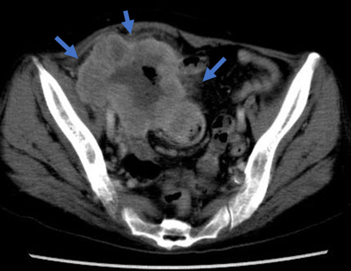
Abdominopelvic contrast-enhanced computed tomography shows a large mass of the cecum and right ovary with well-contrasted circumference vessels.

Abdominopelvic contrast-enhanced computed tomography shows a large mass of the cecum and right ovary with well-contrasted circumference vessels.