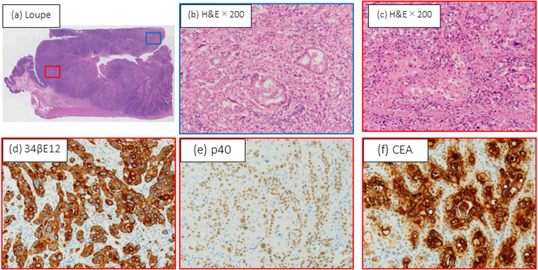
Figure 4.
Microscopic and immunohistochemical features of adenosquamous carcinoma of the cecum. (a) Histological findings. Blue: adenocarcinoma (AD) component; red: squamous cell carcinoma (SCC) component (hematoxylin and eosin [H&E]; loupe). (b) Ductal and cribriform pattern of atypical columnar AD cells (H&E; 200× magnification). (c) Island pattern by dysplastic squamous epithelium with keratinization in SCC component (H&E; 200× magnification). Immunohistochemistry shows SCC component is (d) strongly 34βE12+, (e) p40+, and (f) focally positive for carcinoembryonic antigen ( 200× magnification for each).

