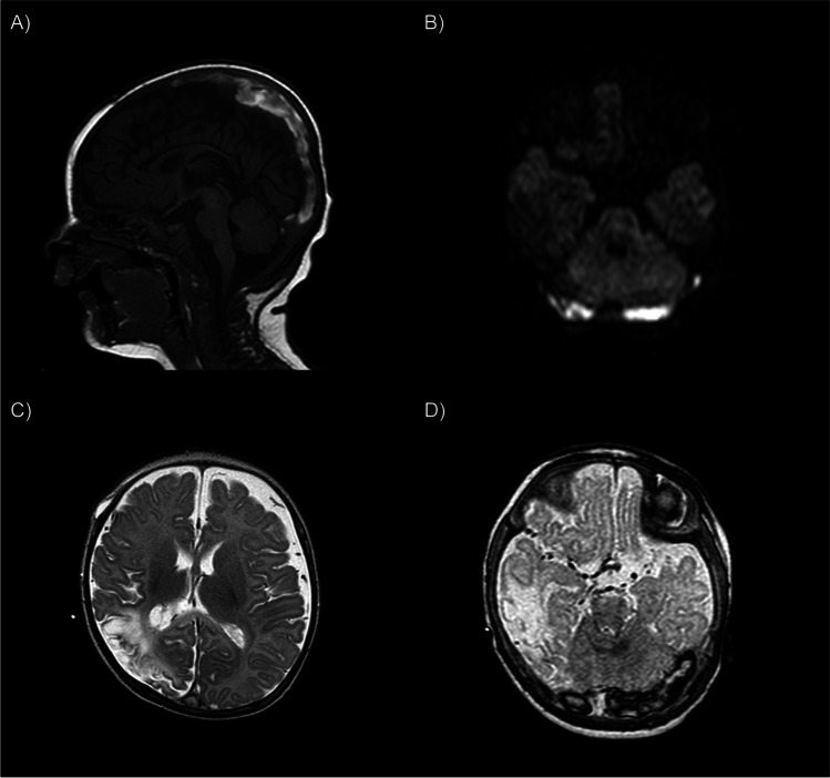
Fig. 2.
MRI done in 2 weeks of the evacuation of the SDH showing CVT. A MRI T1 sag shows the thrombosis of the sagittal sinus and confluens sinuum; B MR diffusion shows the thrombosis invading further into both transverse sinuses; C MRI T2 axial demonstrates the progression of left hemisphere ischemia; D MRI T1W shows the thrombosis of the transverse sinuses

