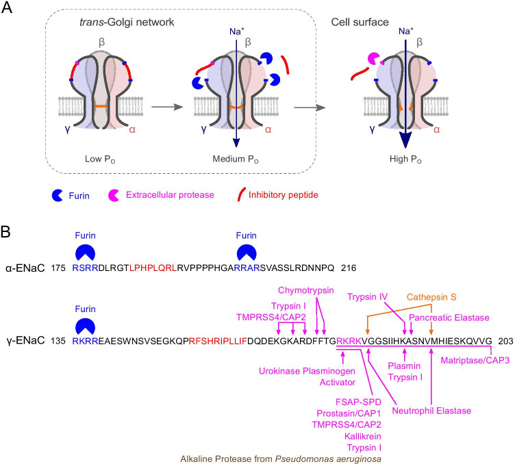
Fig. 1.
ENaC activation by intra- and extracellular proteases. A Cartoon illustration of ENaC cleavage by furin within the trans-Golgi network and extracellular proteases at the cell surface. Please note that αβγ-ENaC assembles in a counter-clockwise subunit orientation [7]. PO, open probability. B Peptide sequences of the α- and γ-subunits of human ENaC showing the regions containing the inhibitory peptides within the extracellular loop. Furin consensus sites are shown in blue; the inhibitory peptides [7] are shown in red. The figure shows extracellular proteases cleaving the γ-subunit whose cleavage sites have been identified by mutagenesis studies [4–6]: Serine proteases are shown in magenta, cysteine proteases in orange, and metalloproteases in brown. CAP, channel-activating protease (alternative name for the indicated proteases); FSAP-SPD, factor VII activating protease—serine protease domain

