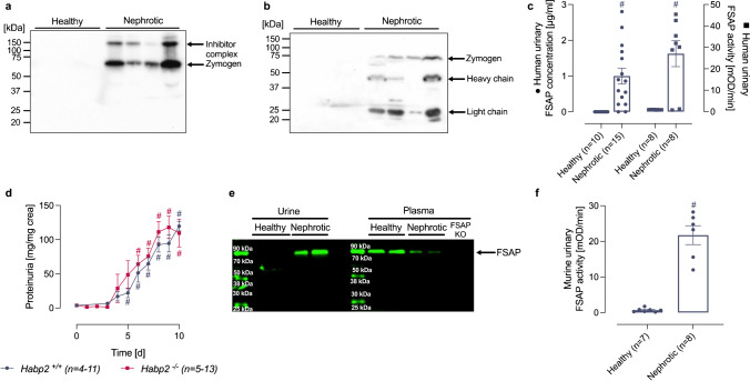
Fig. 1.
Urinary excretion of FSAP in nephrotic syndrome. a Western blot from human urine samples (n = 4 healthy, n = 4 nephrotic) under non-reducing conditions using a rabbit polyclonal antibody. In nephrotic samples, FSAP is detected at 64 kDa as zymogen and at 150 kDa as part of a inhibitor complex. b Western blot of the same samples (n = 4 healthy, n = 4 nephrotic) as used in (A) under reducing conditions using a mix of two monoclonal antibodies. In addition to the detection of FSAP zymogen as single chain (64 kDa), both the light (27 kDa) and heavy chain (50 kDa) are detected which requires previous cleavage at the activation bond R311. Both chains dissociate under reducing conditions. c Quantitation of urinary FSAP concentration and activity in human nephrotic urine samples. Activity was measured with pro-uPA as substrate after immunocapture of FSAP. Results are quantified as uPA chromogenic substrate turnover in mOD min−1. d Proteinuria in wildtype (Habp2+/+) and FSAP-deficient mice (Habp2−/−) after induction of experimental nephrotic syndrome by doxorubicin. e Western blot for FSAP expression from plasma and urine of Habp2+/+ mice (n = 2). FSAP is detected at 64 kDa in its zymogen form in plasma samples and nephrotic urine. Compared to healthy plasma, FSAP expression appears to be reduced most likely due to urinary loss. The antibody does not recognize this band in the plasma from Habp2−/− mice proving the specificity of the antibody. f FSAP activity in mouse urine from Habp2+/+ mice as determined with pro-uPA as substrate. Results are quantified as uPA chromogenic substrate turnover in mOD min−1. #Significant difference between healthy and nephrotic samples

