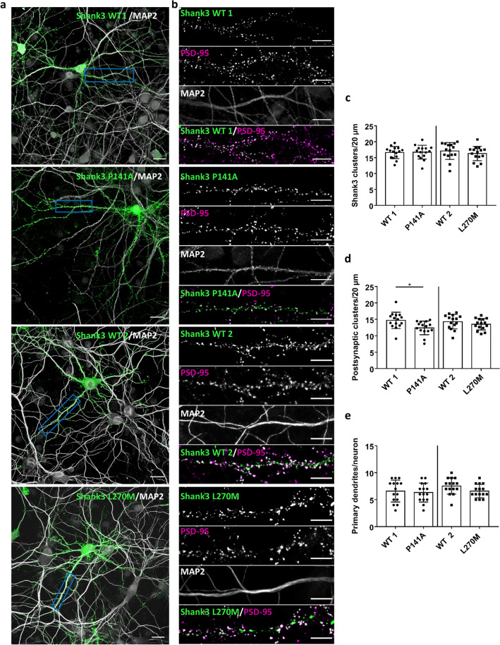
Figure 10.
Primary hippocampal neurons expressing GFP-Shank3 (WT or P141A, L270M mutants) were fixed and stained for the postsynaptic marker PSD-95 and the dendritic marker MAP2 (gray). The expressed Shank3 variants were detected using the GFP fluorescence. (a) Overview images of neurons (scale bar 20 μm). (b) Boxed dendritic areas were magnified (scale bar 10 μm). GFP-Shank3 colocalises extensively with PSD-95 in presumably postsynaptic clusters, which are found in a small distance from the (MAP2-positive) main dendrite. (c) Quantitative evaluation of the total number of Shank3 clusters per 20 μm dendrite. ns: non-significant; analysis of 45 dendrites of n = 15 neurons from three independent experiments; t-Test. (d) Quantitative evaluation of the number of postsynaptic Shank3 clusters (Shank3 clusters colocalising with PSD-95) per 20 µm dendrite. *Significantly different from WT, p < 0.05; analysis of 45 dendrites of n = 15 neurons from three independent experiments; t-Test. (e) Quantitative evaluation of the number of primary dendrites per neuron. ns: non-significant; analysis of n = 15 neurons from three independent experiments; t-Test.

