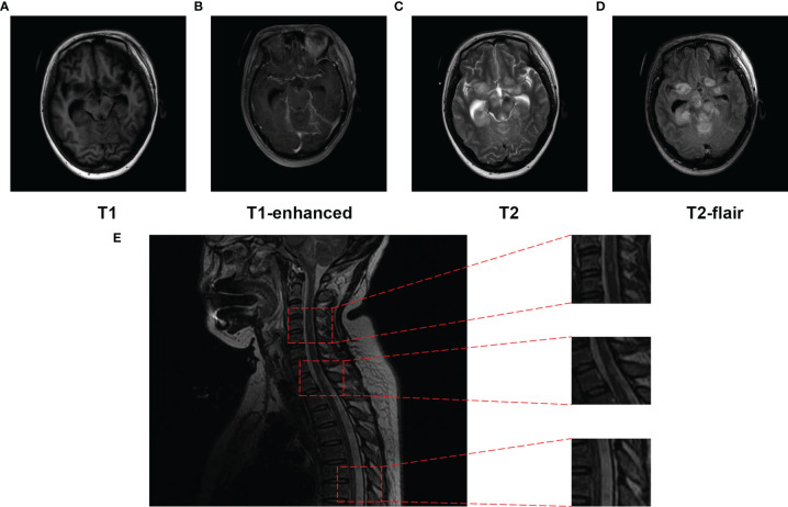
Figure 4.
Magnetic resonance imaging (MRI) of the brain performed and spinal cord (T2) on day 15 post-hospitalization. (A–D) The frontal lobes, temporal lobes, ventriculus lateralis, brainstem, cisterna, and cerebellum were displayed as multiple patches, with high signal on T2WI, low signal on T1WI, apparently high signal on T2-Flair, and T1 enhanced. (E) Punctate and patchy lesions were presented as high T2 signals in the cervical and thoracic spinal cord.

