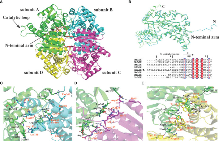
Figure 6.
Subunit interfaces of BoLDH. (A) The tetrameric state of BoLDH. BoLDH consists of four identical subunits, and colored in green, scan, magenta and yellow, respectively. (B) Comparison between apo Bifidobacterium longum (Bl) LDH (1lld) and BoLDH. The apo BoLDH (scan) were superimposed on and BlLDH without ligands (green). (C) The interface of subunit A and subunit B. Carton models of subunits A and B are displayed in different colors (green and scan). (D) The interface of subunit A and subunit C. Carton models of subunits A and C are displayed in different colors (green and magenta). (E) The interface of subunit A and subunit D. Carton models of subunits A and D are displayed in different colors (green and yellow). The dashed lines represent the hydrogen bonds and salt-bridges between the respective donor and acceptor atoms. These pictures were produced using the software PyMOL 4.30.

