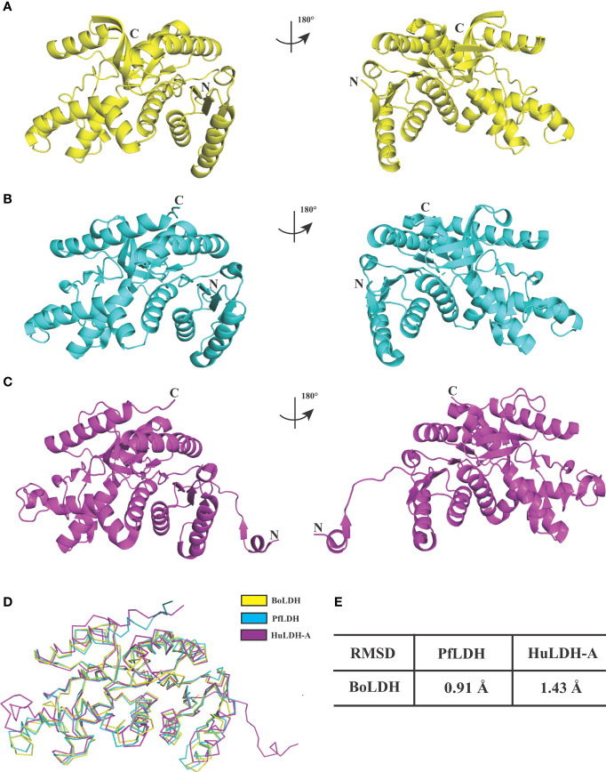
Figure 8.
Structural comparisons of BoLDH, PfLDH and HuLDH-A. (A) Overlay of BoLDH structure at 180 degrees. (B) Overlay of PfLDH structure at 180 degrees (PDB accession number 2x8l). (C) Overlay of HuLDH-A structure at 180 degrees (PDB accession number 4ojn). Structures of the three LDHs were exhibited in an identical orientation as cartoons, and colored in yellow, cyan and purple. (D) Ribbon diagrams of the three LDH structures. (E) Structure identities and RMSD values between BoLDH and PfLDH or between BoLDH and HuLDH-A were shown in the form. The RMSD values were calculated using the PDBeFold service.

