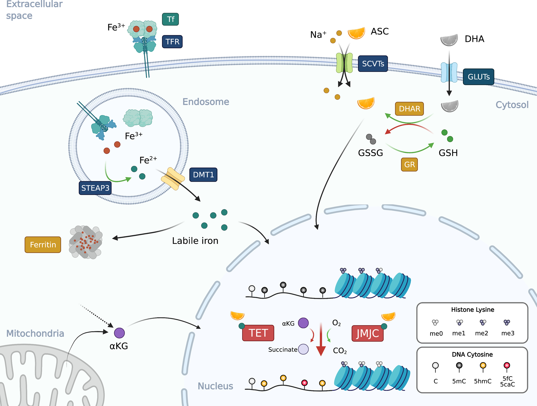
Figure 2. Ascorbate and iron in epigenetic regulation.

Ascorbate (ASC; vitamin C or VC) is co-transported with sodium ion (Na+) from extracellular space to the cytosol via SVCTs (sodium-ascorbate co-transporters) at the ratio of 1-to-2 (ASC to sodium). Dehydroascorbate (DHA), the oxidized form of ASC, enters the cells via the glucose transporters (GLUTs). DHA in the cells is reduced to ASC by dehydroascorbate reductase (DHAR) by oxidizing two molecules of reduced (GSH) into oxidized glutathione (GSSG). GSSG can be reduced back to GSH by glutathione reductase (GR). Iron usually exists in the circulation as the oxidized ferric iron (Fe3+) bound to transferrin (Tf) at a ratio of 2:1. Transferrin receptor (TFR) binds to and internalize the Tf:iron complex to endosome. The ferric iron is released from Tf in the acidic environment and reduced by the metalloreductase STEAP3 (Six-Transmembrane Epithelial Antigen of Prostate 3) into ferrous iron (Fe2+), which then translocates from the endosome to cytosol via DMT1 (Divalent Metal Transporter 1) to form the labile iron pool. Since ferrous iron can catalyze the formation of ROS, its concentration is tightly regulated and is stored as ferric iron in the ferritin complex that consists of 24 subunits. The ferritin complex can hold around 4,500 molecules of Fe3+. Both ASC and ferrous iron are cofactors for 2OGDDs including TET and JMJC proteins that used αKG (2-OG) and molecular oxygen to oxidize the target. TET oxidizes 5mC into 5hmC, 5fC, and 5caC, while various JMJC proteins demethylate histone lysine (and arginine, not depicted). αKG can be derived from mitochondria or cytosol as an intermediate metabolite. Black arrows, movement of molecules; green arrows, reduction; red arrows, oxidation. Blue text boxes represent transmembrane proteins and boxes with other colors indicate the location of the proteins. Figure was created with BioRender.
