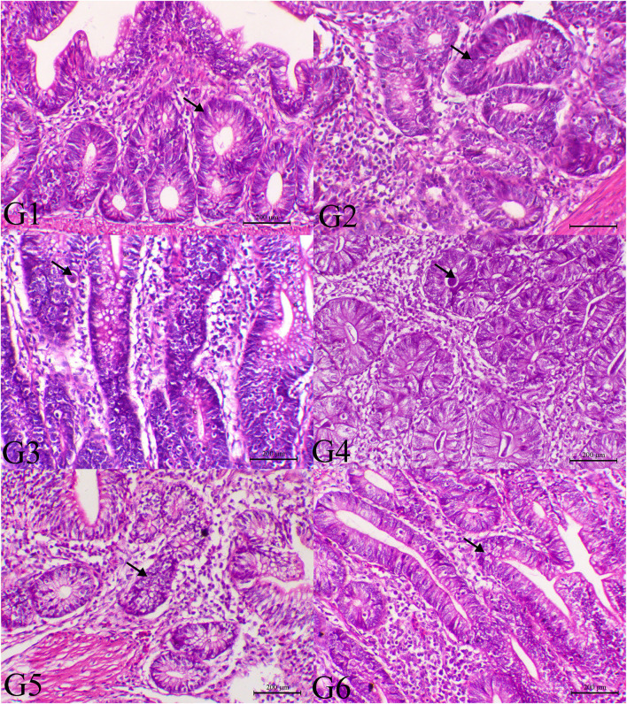
Figure 3.
Histopathology of cecal tissues of the normal bird (G1), SMC-treated birds (G2), E. tenella-challenged birds and examined on the 14th day post infection (G3), challenged and treated birds with SMC low dose (G4), high dose (G5), and diclazuril (G6). G1 and G2 (arrows indicate normal intestinal crypts), G3 (arrow reveals the presence of parasites within the intestinal crypts), G4 (arrow indicates marked decrease of the parasites within the intestinal mucosa), G5 (arrow indicates normal intestinal crypts), and G6 (arrow indicates hyperplastic changes within the intestinal mucosa), H&E stain, bar = 200 μm.

