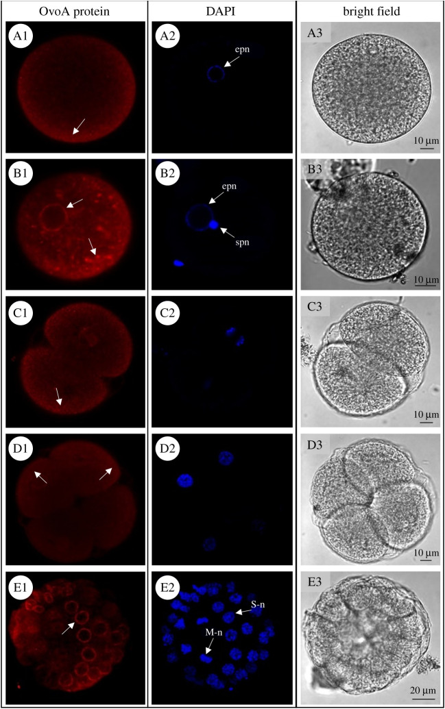
Figure 3.
OvoA spatial expression in P. lividus early developmental stages. For immunohistochemistry experiments (IHC), OvoA immunofluorescence (in red, left side), nuclei (labelled in blue with DAPI, middle) and images in bright field are shown for: (A1–A3) unfertilized eggs; (B1–B3) fertilized eggs; (C1–C3) 2-cell stage; (D1–D3) 4-cell stage; (E1–E3) 32-cell stage. In OvoA protein panel, white arrows indicate OvoA signal (for details refer to the text). epn = egg pronucleus, spn = sperm pronucleus, S-n = S-phase (interphase) nucleus, M-n = M-phase (mitosis) nucleus. Pictures were taken at confocal microscope (Zeiss LSM 700) at 20× magnification.

