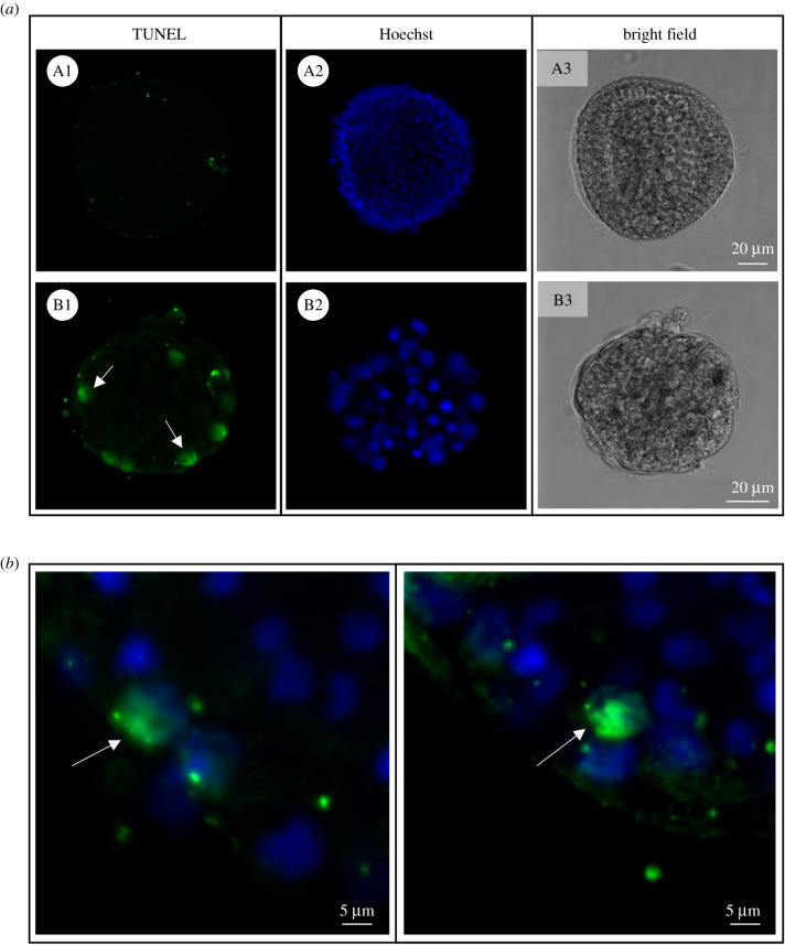
Figure 6.
Apoptotic nuclei detection in OvoA knockdown sea urchin embryos. (a) TUNEL signal, nuclei staining (Hoechst) and bright field images are shown for both ctrl-MASO (A1–A3) and ovoA-MASO embryos (B1–B3). (b) TUNEL/Hoechst merged signal from two additional representative ovoA-MASO embryos. White arrows indicate the TUNEL-positive nuclei.

