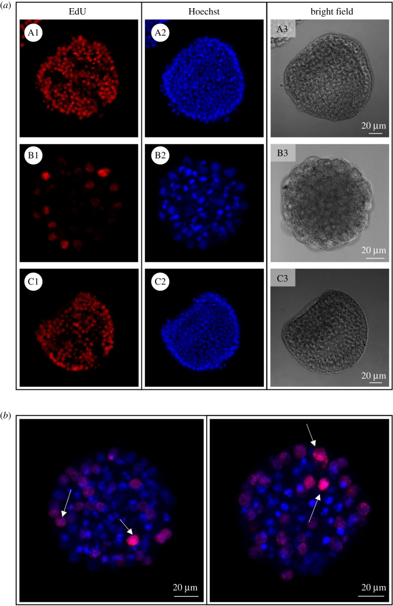
Figure 7.
Proliferating nuclei staining in OvoA knockdown sea urchin embryos. (a) EdU signal, nuclei staining (Hoechst) and bright field images are shown for embryos injected with ctrl-MASO (A1–A3), ovoA-MASO alone (B1–B3) and co-injected with ovoA synthetic mRNA (C1–C3). (b) EdU/Hoechst merged signal from two additional representative ovoA-MASO embryos. White arrows indicate the EdU-positive nuclei. Pictures were taken with a confocal microscope (Zeiss LSM 700) at 20× magnification.

