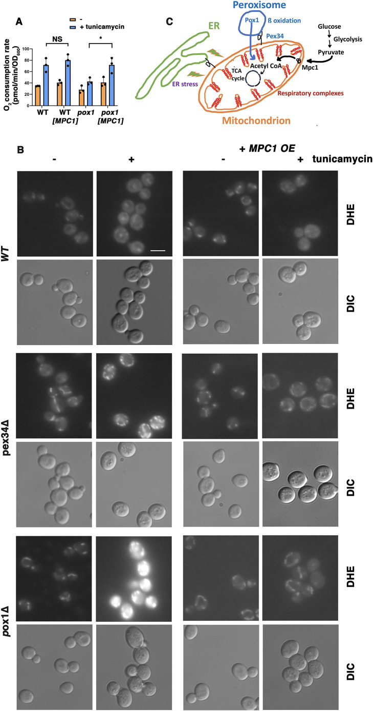
Fig. 3.
Rescue of pox1Δ cells by overexpression of MPC1. (A) OCR measured with or without MPC1 overexpression. OCR of wild-type (WT) cells with and without ERS (0.5 µg/ml tunicamycin for 5 h) was unchanged by MPC1 overexpression; OCR was significantly induced by ERS in wild-type cells. Without MPC1 overexpression, OCR was not significantly increased by ERS in pox1Δ cells. In pox1Δ cells with MPC1 overexpression, OCR was induced significantly by ERS. Data are presented as mean±s.e.m. n=3. *P≤0.05; NS, not significant (Student's t-test, one-tailed, unpaired). (B) ROS accumulation in pox1Δ and pex34Δ cells is ameliorated by MPC1 overexpression. WT and mutant cells with or without a HIS3-marked high-copy plasmid bearing MPC1 (MPC1 OE) were treated with or without tunicamycin (0.5 µg/ml tunicamycin for 5 h) and then stained with DHE to detect ROS, as described for Fig. 2A. Fluorescence images were collected at the same exposure and adjusted with Photoshop using the same settings. Scale bar: 10 µm. Images are representative of three experiments. (C) Schematic diagram illustrating the mitochondrial respiratory response to ERS and the requirement for fatty acid β-oxidation contributed by peroxisomes and pyruvate delivered to mitochondria via the Mpc1 carrier.

