Fig. 3.
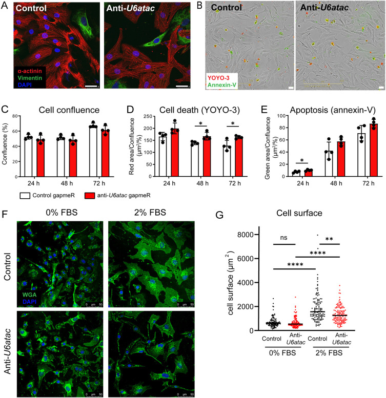
Impact of minor splicing knock-down on cardiomyocyte growth, survival and size. (A) Representative immunocytochemistry of three experiments of NRVMs at 48 h after transfection with control or anti-U6atac gapmeRs. Red, α-actinin (cardiomyocyte marker); green, vimentin (fibroblast marker); blue, DAPI. (B) Representative phase-contrast images of NRVMs and (C) quantification of cell confluence, (D) cell death and (E) apoptosis. Data are presented as mean±s.d. for 4 wells per condition. *P<0.05 (unpaired two-tailed Student's t-test). (F) Representative images of transfected NRVMs after 48 h incubation in medium with 0% FBS or 2% FBS and (G) quantification. Green, WGA (membrane marker); blue, DAPI. n>100 cells per condition. **P<0.01; ****P<0.0001; ns, not significant [one-way ANOVA on ranks (Kruskal–Wallis test), followed by Dunn's test for post hoc analyses]. Scale bars: 25 µm (A,B); 50 µm (F).

