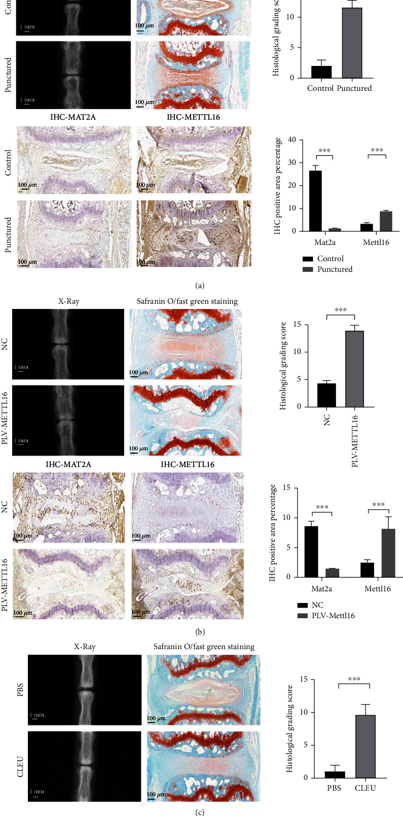
Figure 6.

In vivo studies. (a) Verification of the disc degeneration animal model. X-ray examination revealed a significant decrease of the height of the punctured disc. Safranine O/fast green staining showed that the punctured NP tissue became more disorganized, and fewer cells could be seen. The histological grading score was significantly higher in the punctured disc. Immunohistochemical analysis demonstrated less MAT2A protein and more METTL16 protein levels in the punctured NP tissues. (b) Degenerative changes in the discs injected with METTL16 overexpression lentivirus. X-ray examination revealed significant decrease of the height of the discs. Safranine O/fast green staining showed that the NP tissue became more disorganized, and fewer cells could be seen. The histological grading score was also significantly higher. Immunohistochemical analysis demonstrated less MAT2A protein and more METTL16 protein levels in the NP tissues. (c) Degenerative changes in the discs injected with cycloleucine. X-ray examination revealed significant decrease of the height of the discs. Safranine O/fast green staining showed that the NP tissue became more disorganized, and fewer cells could be seen. The histological grading score was also significantly higher. n = 3 replicates per group, ∗∗∗p < 0.001, 0.001 ≤ ∗∗p < 0.05, ∗p < 0.05.
