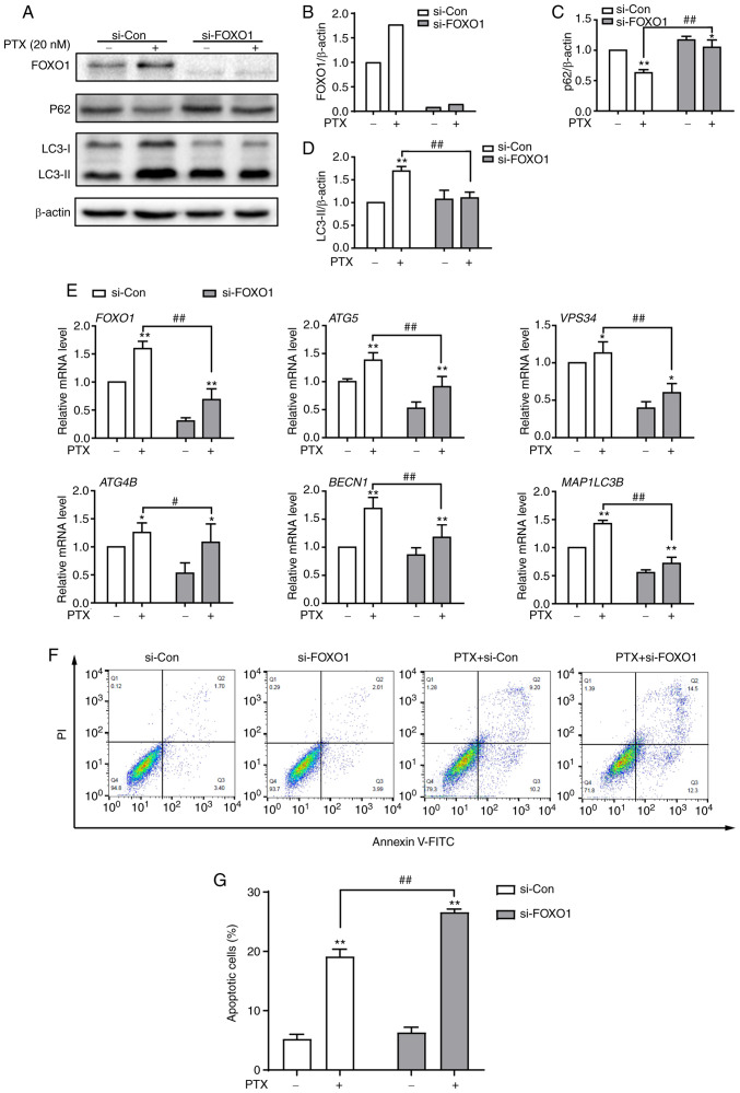
Figure 6.
Knockdown of FOXO1 attenuates PTX-induced autophagy and promotes apoptotic cell death in MDA-MB-231 cells. (A) Protein expression of the autophagy markers P62, LC3-I and LC3-II after treatment with si-FOXO1, with β-actin as an internal control. Semi-quantification of (B) FOXO1, (C) P62 and (D) LC3-II protein expression. (E) The mRNA expression levels of FOXO1 and its downstream target genes after treatment with siRNA and/or PTX. (F) The effect of FOXO1 knockdown on PTX-induced apoptosis was analyzed by flow cytometry. (G) Quantification of the ratio of apoptotic cells after the knockdown of FOXO1. PTX was used at a concentration of 20 nM. Data are presented as the means ± SD of three independent experiments. *P<0.05 vs. 0 nM PTX; **P<0.01 vs. 0 nM PTX; #P<0.05 vs. 20 nM PTX + si-Con; ##P<0.01 vs. 20 nM PTX + si-Con. PTX, paclitaxel; FOXO1, forkhead box transcription factor O1; LC3, light chain 3; si, small interfering; ATG5, autophagy related 5; VPS34, class III phosphoinositide 3-kinase vacuolar protein sorting 34; ATG4B, autophagy related 4B cysteine peptidase; BECN1, beclin 1; MAP1LC3B, microtubule associated protein 1 light chain 3β.

