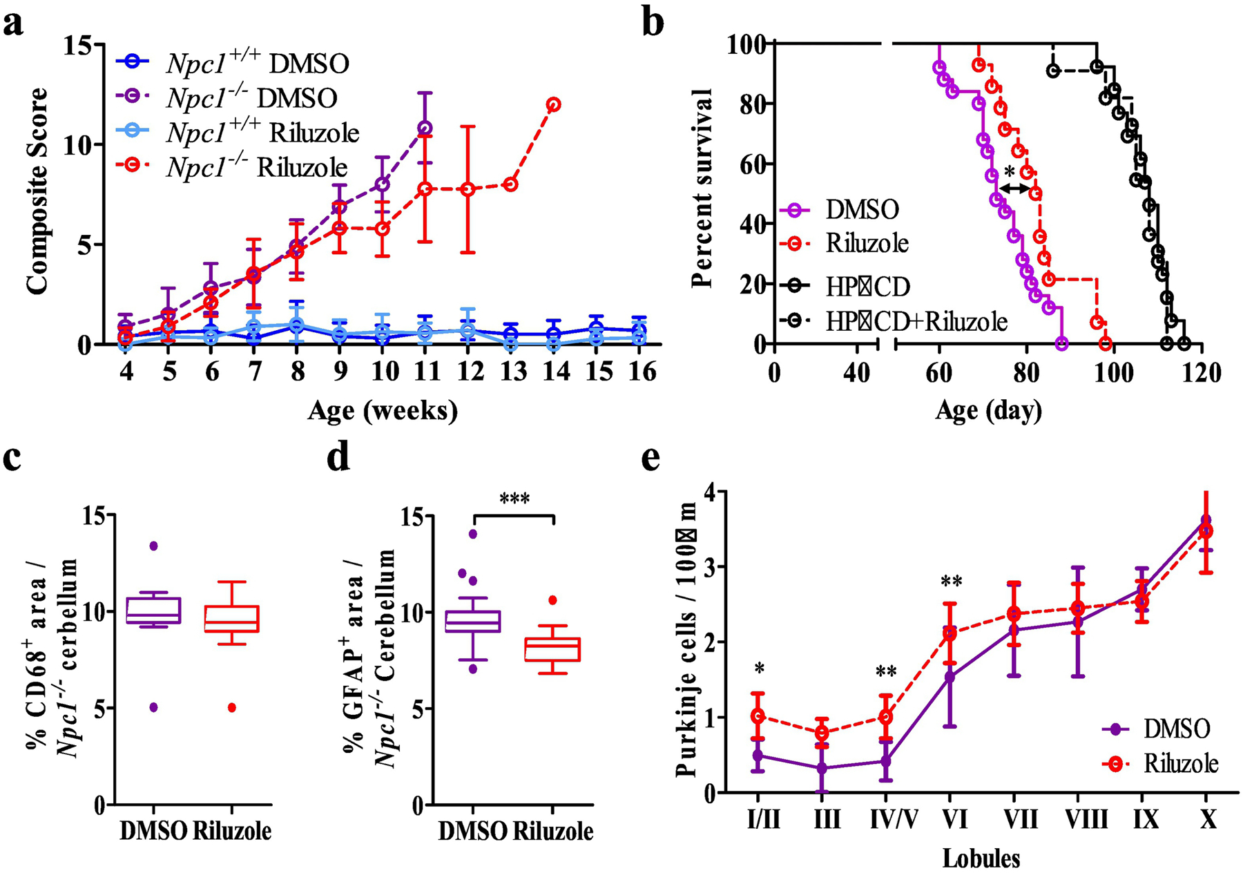
Figure 4.

In vivo evaluation of riluzole. a NPC1 disease composite score evaluation between 4 and 16 weeks of age with DMSO (initial n=16 and 14) and riluzole treated (60µg.ml−1 in the drinking water, initial n=12 and 18) Npc1−/− and Npc1+/+ mice. Initial n values are for Npc1−/− and Npc1+/+ respectively). These numbers become truncated as the experiment progresses. b Kaplan-Meier survival analysis of DMSO (n=20), riluzole (n=14), HPβCD (n=13) and HPβCD + riluzole (n=12) treated Npc1−/− mice. c Immunofluorescence quantification of cerebellar CD68 staining in 7-week-old of DMSO or riluzole treated Npc1−/− mice. d Immunofluorescence quantification of cerebellar GFAP staining in 7-week-old of DMSO or riluzole treated Npc1−/− mice. e Cerebellar lobule Purkinje cell density per 100µm in 7-week-old of PBS or ceftriaxone treated Npc1−/− mice. Purkinje neurons were identified by immunostaining for calbindin. N≥6, Npc1−/− DMSO (purple), Npc1−/− riluzole (red), Npc1+/+ DMSO (blue), Npc1+/+ Riluzole (light blue). *p<0.05, **p<0.01 and ***p<0.001.
