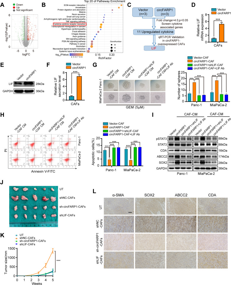
Fig. 3.
circFARP1 enhances the expression and secretion of LIF in CAFs. A Plot showing the sums of the expression levels of genes regulated by circFARP1. B Top 20 enriched pathways of differential mRNA expression between CAFs and circFRAP1-overexpressing CAFs. C Flow chart for the identification of LIF as the downstream target of circFARP1. D-F The mRNA level (D), protein level (E), and secretion level (F) of LIF in CAFs transfected with vector or circFARP1. G-I Panc-1 and MiaPaCa-2 cells were grown in conditioned medium (CM) from CAFs transfected with empty vector or circFARP1 for 2 weeks and subjected to the indicated experiments. Lenti-LIF shRNA or a neutralizing antibody against LIF was used to deplete LIF in CAF-CM. (G) Sphere formation assays of Panc-1 and MiaPaCa-2 cells with the indicated treatments. Scale bars, 50 μm. (H) Flow cytometry analysis of GEM-induced (10 μM) apoptosis in Panc-1 and MiaPaCa-2 cells with the indicated treatment. (I) western blot analysis of pstat3/stat3, ABCC2, CDA, and SOX2 protein expression in Panc-1 and MiaPaCa-2 cells with the indicated treatment. J-L Panc-1 cells were subcutaneously coinjected with or without CAFs stably transfected with lenti-NC-shRNA, lenti-circFARP1-shRNA or lenti-LIF-shRNA into nude mice followed by GEM treatment (50 mg/kg). UT, untreated Panc-1. J Representative images of xenograft tumors of each group. K Tumor growth curve were shown. L Representative images of IHC for α-SMA, SOX2, ABCC2, and CDA in xenograft tumors. Scale bars, 50 μm. Data are expressed as the mean ± SD. ***p<0.001

