
Extended Data Fig. 5 |. ppil4−/− zebrafish exhibit apoptosis in neurons and radial glia, but not in endothelial cells.
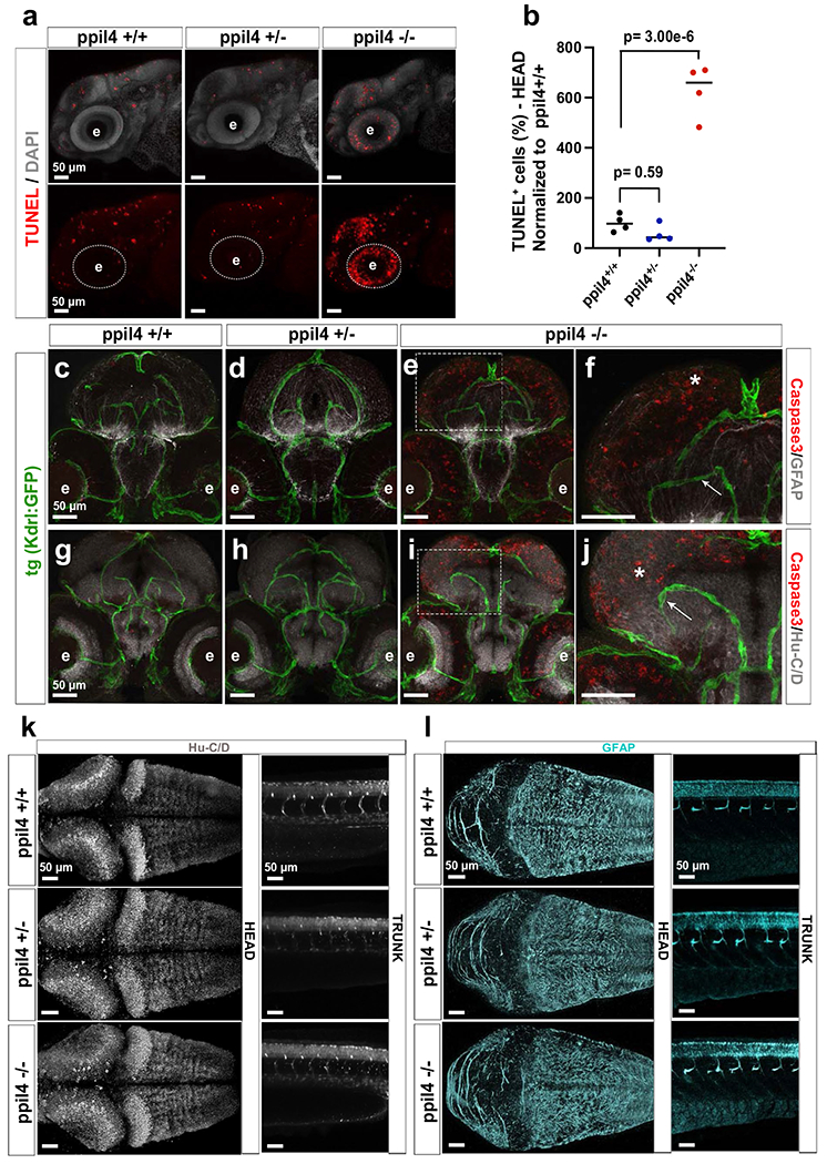
a,b, Representative confocal images of 2.5 dpf embryos, where ppil4−/− mutants exhibit an increase in TUNEL-positive cells in the head region, n=4 sets biological replicates with 30 zebrafish per set. c-j, Cross-sections of the head. Embryos at 2.5 dpf in tg(kdrl:gfp)zn1 background were stained for Caspase-3 and HU (neurons) or GFAP (radial glia). Apoptosis was detected in neurons and radial glia (white asterisks), but not in endothelial cells (white box; arrows). Confocal images, n= 3 sets biological replicates with 30 zebrafish per set. k,l, Whole mount confocal images of 2.5 dpf embryos showing no difference in (k) neuronal (HU+) and (l) radial glial (GFAP+) population in ppil4 mutant genotypes, n= 3 sets biological replicates with 30 zebrafish per set. Dorsal view of the head region (left panel), lateral view of the trunk (right panel). Individual values shown with scatter dot plot and median. Statistical test: (b) One-way ANOVA followed by Dunnett’s multiple comparison test. Abbreviations: TUNEL: Terminal deoxynucleotidyl transferase dUTP nick end labeling; Fb: forebrain; Mb: midbrain; Hb: hindbrain; e=eye. Scale bar: 50 μm.