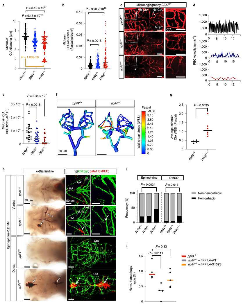
Fig. 3 |. Depletion of ppil4 leads to cerebral hemorrhage.

a, Midbrain CtA diameter in ppil4+/+ (n = 153), ppil4+/− (n = 147) and ppil4−/− (n = 140) embryos at 2.5 dpf. Orange bar: Levene’s test. b, Vascular resistance in midbrain CtAs in ppil4+/+ (n = 143), ppil4+/− (n = 129) and ppil4−/− (n = 109) embryos at 2.5 dpf. c, Microangiography by transcardiac injection of BSA594 (2.5 dpf), n = 10 per genotype. d,e, Representative time–velocity plots (d) and comparison of blood flow (e) in midbrain CtAs at 2.5 dpf, n = 27, 26 and 33 vessels in ppil4+/+, ppil4+/− and ppil4−/−, respectively. f,g, Wall shear stress in 3D cerebrovascular models of ppil4+/+ (n = 4) and ppil4−/− (n = 6) zebrafish (2.5 dpf) using ANSYS. h, Bright-field (o-Dianisidine staining) and confocal images of tg(kdrl:GFP;gata1:dsRED) of ppil4+/+ (n = 70) and ppil4−/− zebrafish (n = 78) embryos treated with epinephrine (AA1 and AA2, first and second aortic arch arteries; HA, hypobranchial artery). i, Hemorrhagic events in brain and aortic arch arteries after epinephrine (h) versus DMSO (vehicle) treatment at 72 hpf, nepi = 70, 163 and 78 and nDMSO = 56, 121 and 66 for ppil4+/+, ppil4+/− and ppil4−/−, respectively. j, Normalized frequency of epinephrine-induced hemorrhage in brain and aortic arch arteries in uninjected, hPPIL4WT- or hPPIL4G132S-injected ppil4−/− embryos, n = 4, 4 and 5 sets of biological replicates with 200 zebrafish per set. Individual values are shown with scatter plot and median in a,b,e,g and j. Statistical tests: Kruskal-Wallis test followed by Dunn’s test (a,b); Levene’s test (based on median) (a); one-way ANOVA with Dunnett’s multiple comparison test (e,j); two-tailed Mann-Whitney test (g); pairwise Fisher’s exact test with FDR correction (i). Scale bar, 50 μm. WSS, wall shear stress.
