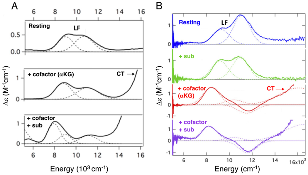
Figure 2:

Near IR MCD spectra of FeII sites in FIH (A) and DAOCS (B) for resting enzyme (top), substrate bound (right second down), akg bound (left middle and right third down) and both akg and substrate bound (bottom). Ferrous ligand field (LF) transitions and charge transfer (CT) transitions. Figure adapted from [Iyer, S. R. et al. (2018) O2 activation by non-heme FeII α-ketoglutarate-dependent enzyme variants: Elucidating the role of the facial triad carboxylate in FIH. Journal of the American Chemical Society 140, 11777–11783.] and [Goudarzi, S. et al. (2020) Evaluation of a concerted vs. sequential oxygen activation mechanism in α-ketoglutarate—dependent nonheme ferrous enzymes. Proceedings of the National Academy of Sciences 117, 5152–5159]. Copyright [2018] and [2020] respectively.
