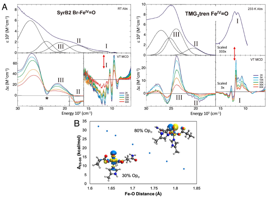
Figure 8:

Experimental insight into dπ* FMO. A. Abs (top) and MCD (bottom) definition of FeIV=O FMOs in SyrB2 (left) and its TMG3tren model complex (right). Red double arrows indicate dπ* to dσ* LF excitation at an Fe=O bond length of 1.62 Å. B. Dependence of the dπ* to dσ* excitation energy (the promotion energy (PE) in Fig. 7B) on Fe=O bond length (1.82 Å at the TS). Insets show the σ and π FMOS at an Fe=O distance of 1.62 Å. Adapted from [Srnec, M. et al. (2020) Nuclear Resonance Vibrational Spectroscopic Definition of the Facial Triad FeIV=O Intermediate in Taurine Dioxygenase: Evaluation of Structural Contributions to Hydrogen Atom Abstraction. Journal of the American Chemical Society 142, 18886–18896.] and [Srnec, M. et al. (2016) Electronic Structure of the Ferryl Intermediate in the alpha-Ketoglutarate Dependent Non-Heme Iron Halogenase SyrB2: Contributions to H Atom Abstraction Reactivity. Journal of the American Chemical Society 138, 5110–5122.] Copyright [2020] and [2016] respectively.
