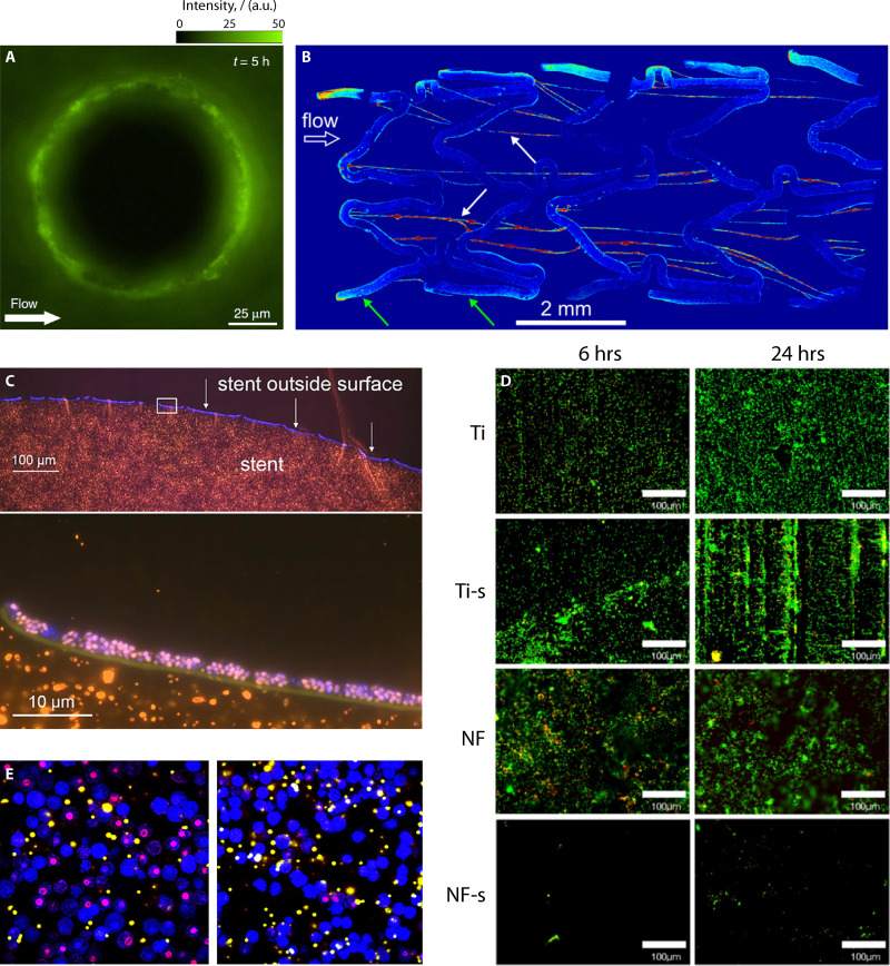
FIG 3.
Mosaic of representative images. (A) Image of fluorescent P. aeruginosa bacteria attached to a 100-μm pillar in the presence of flow. Modified from Secchi et al. (289). (B) Maximum intensity projection image of biofilm streamers formed flowing a suspension of fluorescent P. aeruginosa in a bare-metal stent. Modified from Drescher et al. (312). (C) Bacterial colonization of a biliary polyethylene stent in a patient with bile duct stenosis, visualized by fluorescence in situ hybridization (FISH). Modified from Yan and Bassler (92). (D) Fluorescence images of E. coli on titanium (Ti), silanized titanium (Ti-s), nanoflower-coated (NF), and nanoflower-coated surfaces after silanization (NF-s). Modified from Montgomerie and Poat (333) with permission of Elsevier. (E) Confocal images (X63 magnification) of bone marrow-derived macrophage phagocytosis of fluorescent microspheres (yellow-white) and cell death with propidium iodide stain (red-purple) after exposure to S. aureus biofilm-conditioned medium (left-hand side) and S. aureus planktonic culture-conditioned medium (right-hand side). Modified from Torres et al. (383).

