. 2021 Sep 28;13(3):595–610. doi: 10.1039/d1sc03327a

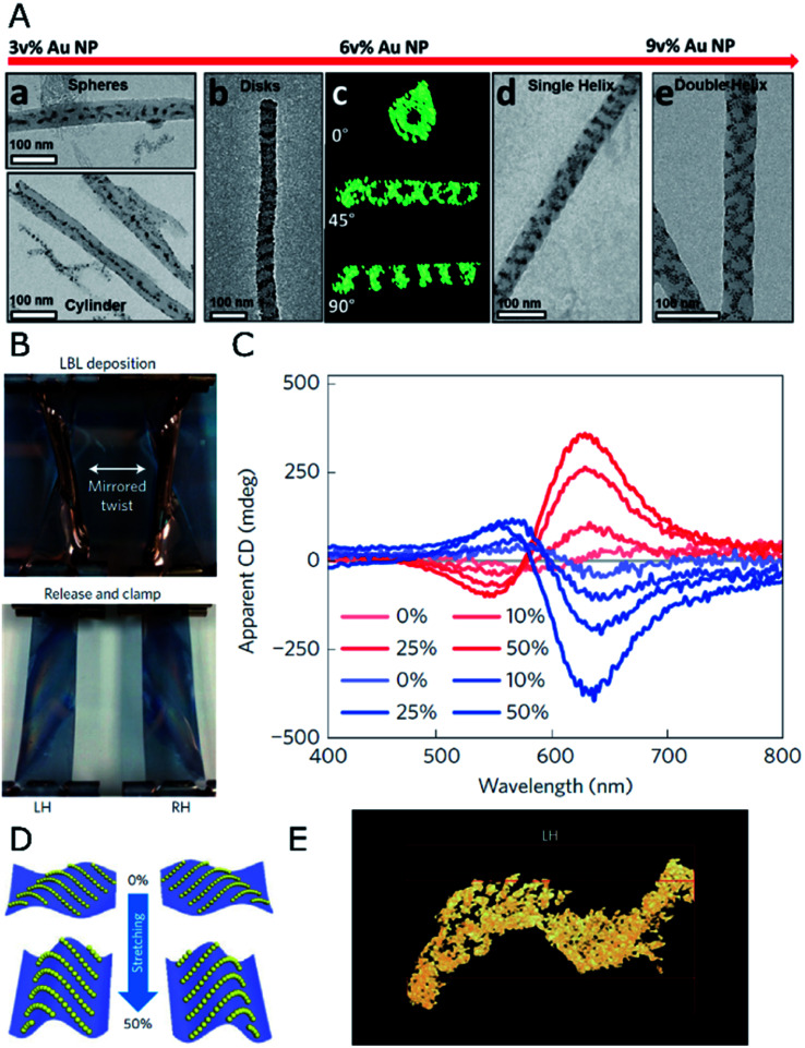
Fig. 1. (A) TEM and electron tomography of BCP-Au NP structures obtained for different NP loadings through co-assembly under confinement in AAO nanopores, displaying spherical and 1D arrays at 3 vol%, disks and single- or double-helices at 6 and 9 vol%, as labeled. Reproduced from ref. 59 with permission from American Chemical Society, copyright 2017. (B) Photographs of twisted, clamped PDMS substrates with opposite handedness after poly(urethane) and Au NP layer-by-layer deposition, and subsequent relaxation back into the planar state. (C) Apparent CD spectra for both (L- and R-, red and blue curves respectively) samples under different stretching percentages. (D) Scheme of NP arrangement in both samples under 0% and 50% stretching. (E) TEM tomograph showing the S-like surface topography of the cross-sectioned left-handed sample. (B–E) Reproduced from ref. 62 with permission from Nature Publishing Group, copyright 2016.

Fig. 1