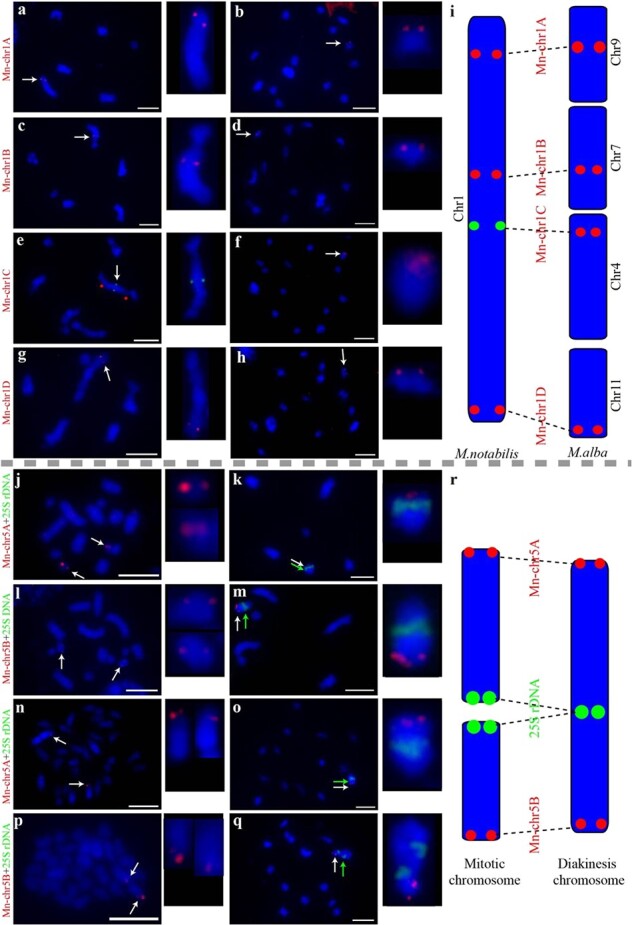
Figure 3.

Chromosomal collinearity patterns verified by FISH using single-copy sequence probes and a 25S rDNA probe in M. notabilis and M. alba ‘Lunjiao109’. Probe Mn-chr1A mapped to the upper distal part of diakinesis chromosome 1 of M. notabilis (a) and diakinesis chromosome 9 of ‘Lunjiao109’ (b). Probe Mn-chr1B mapped to the middle part of diakinesis chromosome 1 of M. notabilis (c) and diakinesis chromosome 7 of ‘Lunjiao109’ (d). Probe Mn-chr1C mapped to the middle part of diakinesis chromosome 1 of M. notabilis. Red stars mark two clear chromosome constrictions on chromosome 1 (e). Probe Mn-chr1C mapped to diakinesis chromosome 4 of ‘Lunjiao109’ (f). Probe Mn-chr1D mapped to the tail segment of diakinesis chromosome 1 of M. notabilis (g) and diakinesis chromosome 11 of ‘Lunjiao109’ (h). Chromosomes were stained with DAPI (blue). All probe signals and the chromosomes where they were located are shown to the right in each cell at greater magnification. Arrows indicate the FISH signals of the probes. Scale bars represent 5 μm. Schematic representation of the collinearity pattern between chromosome 1 of M. notabilis and chromosomes 4, 7, 9, and 11 of M. alba. Probe names are shown in red font (i). Probe Mn-chr5A mapped to the smallest mitotic chromosome (chromosome 7) pair of M. notabilis (j) and the terminal region of the short arm of diakinesis chromosome 5 of M. notabilis, which was indicated by the centrally located signal of 25S rDNA (indicated by a green arrow) (k). Probe Mn-chr5B mapped to the mitotic chromosome 5 pair of M. notabilis (l) and the terminal region of the long arm of diakinesis chromosome 5 of M. notabilis, which was indicated by the centrally located signal of 25S rDNA (indicated by a green arrow) (m). Probe Mn-chr5A mapped to the terminal region of one pair of the mitotic chromosomes of ‘Lunjiao109’ (n) and the terminal region of the long arm of diakinesis chromosome 1 of ‘Lunjiao109’, which was indicated by the centrally located signal of 25S rDNA (indicated by a green arrow) (o). Probe Mn-chr5B mapped to the terminal region of one pair of mitotic chromosomes of ‘Lunjiao109’ (p) and the terminal region of the short arm of diakinesis chromosome 1 of ‘Lunjiao109’, which was indicated by the centrally located signal of 25S rDNA (indicated by a green arrow) (q). Chromosomes were stained with DAPI (blue). All probe signals and the located chromosomes are shown to the right in each cell at greater magnification. Arrows indicated the FISH signals of the probes. Scale bars represent 5 μm. Schematic representation of the chromosomal fusion events from mitotic chromosomes to meiotic chromosomes in M. notabilis and M. alba. Probe names are shown in red font (r).
