. 2021 Jun 17;8(1):281–286. doi: 10.2176/nmccrj.cr.2020-0300

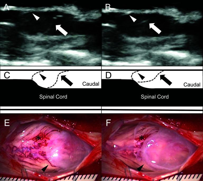
Fig. 2. Intraoperative ultrasonograms (A, B) with the illustrations (C, D), and photos (E, F) around C7. Two membranes and an indentation of the spinal cord are observed on intraoperative ultrasonograms (AD): both the cranial membrane (arrowhead) and the caudal membrane (arrow) are continuously fluttering, but the movement of the cranial membrane is smaller, suggesting that the cranial membrane is tighter than the caudal membrane. After cutting the arachnoid membrane rostrally to the cranial membrane (arrowhead) and flowing out the CSF, the cranial membrane remains bulging and pulsating: note the relationships between the cranial membrane and the small vessels on the cord (asterisk) (E, F). CSF: cerebrospinal fluid.

Fig. 2