. 2021 Dec 22;8(1):817–825. doi: 10.2176/nmccrj.cr.2021-0268

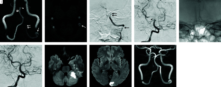
Fig. 1. Case 1. (a) Initial MRA. An abnormal hyperintense signal suggesting intramural thrombus is observed around the transposition part between the left V3 and V4 segments (arrow). The signal of the left VA is faint and interrupted at the V4 segment (double arrow). The top of the BA including the left SCA and the bilateral PCA is not depicted (arrowhead). Only the right SCA is preserved (double arrowheads). (b) Source image of initial MRA. The intimal flap and double lumen are demonstrated at the end of the V3 segment of the left VA (arrow). (c) Initial left vertebral angiogram. The left VA exhibits irregular long segment stenosis at the V3 segment (arrow), followed by irregular tapered occlusion (double arrow). (d) Initial right vertebral angiogram. A filling defect indicating a large clot (arrowhead) extends from the top of the BA to the left SCA and bilateral PCA. (e) Snapshot image of Penumbra ACE68. Penumbra ACE68 has already reached the occlusion site of the BA via the right VA. Afterward, thrombectomy by contact aspiration was performed. (f) Right vertebral angiogram after first pass of contact aspiration. Recanalization of the top of the BA is confirmed. (g and h) DWI obtained a day after the treatment. Small infarcts are observed in the territory of the left AICA and left PCA. (i) MRA obtained 6 months after the treatment. Left VA is spontaneously recanalized without any remaining findings of dissection. MRA: magnetic resonance angiography, VA: vertebral artery, BA: basilar artery, SCA: superior cerebellar artery, PCA: posterior cerebral artery, DWI: diffusion-weighted imaging, AICA: anteroinferior cerebellar artery.

Fig. 1