. 2021 Dec 22;8(1):817–825. doi: 10.2176/nmccrj.cr.2021-0268

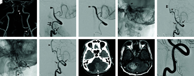
Fig. 3. Case 3. (a) Initial CTA. CTA shows a long, irregular stenosis of the left V3 segment (arrow), a contrast filling defect extending from the left V4 segment to the proximal BA (arrow heads), and occlusion of the right VA. The distal part of the BA is depicted with the collateral flow via the bilateral posterior communicating artery. (b) Initial left vertebral angiogram (lateral view). The intimal flap (arrow) followed by a long irregular stenosis (double arrow) is shown at the left V3 segment. The left VA is interrupted at the distal part of the V4 segment (arrowhead). (c) Superselective injection via the microcatheter (lateral view). A crescent-shaped filling defect suggesting embolic occlusion is demonstrated at the distal end of the V4 segment (arrowhead). (d) Mechanical thrombectomy for left V4 segment occlusion. Trevo XP 4 × 20 mm is deployed from the proximal BA to the distal left V4 segment (arrowhead). (e) Left vertebral angiogram after thrombectomy (lateral view). Although the left V4 segment is recanalized (arrowhead), blood flow to the BA is still restricted (double arrowhead) by severe stenosis of the left V3 segment resulting from dissection (arrows). (f) Left vertebral angiogram after the stenting (lateral view). Two Enterprise 2 VRDs were deployed at the dissected V3 segment in an overlapping manner. Markers of the distal stent (arrows) and proximal stent (arrowheads) are shown. (g) Left vertebral angiogram after the stenting (lateral view). Good dilatation of the left V3 segment (arrows) and complete restoration of blood flow to the BA (arrowhead) are confirmed. (h) CT immediately after the decompression craniectomy. The infarction and oedema in the right cerebellar hemisphere are observed. (i) FLAIR–MRI obtained 30 days after the treatment. The haemorrhagic infarction in the territory of the right PICA and AICA is revealed. (j) Left vertebral angiogram obtained 12 months after stenting (lateral view). The morphological normalization of the left V3 segment with no in-stent restenosis is demonstrated. CTA: computed tomography angiography, BA: basilar artery, VA: vertebral artery, VRD: Vascular Reconstruction Device, CT: computed tomography, FLAIR: fluid-attenuated inversion recovery, MRI: magnetic resonance imaging, AICA: anteroinferior cerebellar artery, PICA: posteroinfeior cerebellar artery.

Fig. 3