. 2021 Nov 19;8(1):787–792. doi: 10.2176/nmccrj.cr.2021-0248

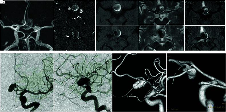
Fig. 1. (A–E) The MRI scans 20 days prior to the first coil embolization indicate a signal void on the right side of the mass (A, B1, B2; TOF-MRA), surrounded by iso-intensity of the center and high-intensity peripherally of the T1-weighted images (C1, C2), heterogeneity intensity of the T2-weighted images (D1, D2), and fluid-attenuated inversion recovery (E1, E2). (F, G) Right CAG performed 15 days prior to the first coil embolization. (F) anteroposterior view, (G) left lateral view, (H) left CAG (3D DSA), left anterior-oblique view, (I) Right CAG (3D DSA), left posterior-oblique view. Acom: anterior communicating artery, CAG: carotid angiography, MRI: magnetic resonance imaging, TOF-MRA: time-of-flight magnetic resonance angiography, 3D DSA: three-dimensional digital subtraction angiography.

Fig. 1