. 2021 Oct 16;8(1):681–690. doi: 10.2176/nmccrj.cr.2021-0107

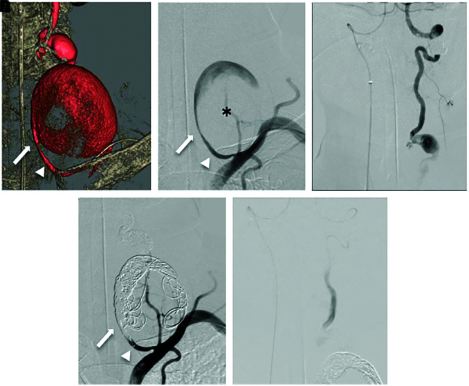
Fig. 2. Three-dimensional digital subtraction angiography (A) showing two fusiform aneurysms located in the V1 to V2 segments of the left VA (proximal aneurysm: thrombosis [asterisk] with MaxD 5.8 mm, distal aneurysm: no thrombus with MaxD 14 mm). Left VAG (B) showing no findings of VA dissection and leakage of the contrast medium. Right VAG (C) showing retrograde blood supply to the left PICA and the aneurysm in the left V2 segment beyond the VA union. Bilateral VAG (D and E) showing complete obliteration of the aneurysm. The arrow: the inflow zone of the proximal giant aneurysm, the arrowhead: VA. MaxD: maximum diameter, PICA: posterior inferior cerebellar artery, VA: vertebral artery, VAG: vertebral angiography.

Fig. 2