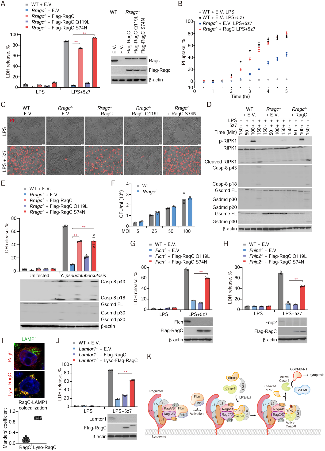
Fig. 6. GTPase activity of lysosomal membrane-anchored RagC is required for caspase-8 activation and pyroptosis triggered by LPS/5z7 or pathogenic Yersinia.

(A, B, C and E) The indicated iBMDMs were treated with LPS, LPS/5z7 or Yersinia. Cell death was measured by LDH release after 2.5 hr (A) or 5 hr (E), or by entry of PI into cells in real-time (B), or as observed by phase-contrast fluorescence microscopy (C). (D) Full-length and cleaved products of caspase-8, RIPK1, Gsdmd and Gsdme from whole-cell lysates of the indicated iBMDMs treated with LPS or LPS/5z7 for the indicated times. (F) The number of Yersinia taken up by WT or Rragc−/− iBMDMs was quantified by counting CFU. (G, H and J) WT, Flcn−/−, Fnip2−/− or Lamtor1−/− iBMDMs reintroduced with the indicated plasmids were treated with LPS/5z7 and assayed for LDH release 2.5 hr post treatment. (I) Lysosomal localization of ectopically expressed RagC and Lyso-RagC in Lamtor1−/− iBMDMs was assessed by confocal microscopy (representative images, upper panel). Colocalization of RagC with LAMP1 was analyzed by calculating Manders’ overlap coefficient (lower panel). (K) Model of RIPK1-caspase-8 activation mediated by Rag-Ragulator complex. Graphs in A, B, E-H, and J show mean ± SEM of triplicate wells. Data are representative of at least three independent experiments. Data were analyzed using a two-tailed Student’s t test. **P < 0.01. E.V., empty vector.
