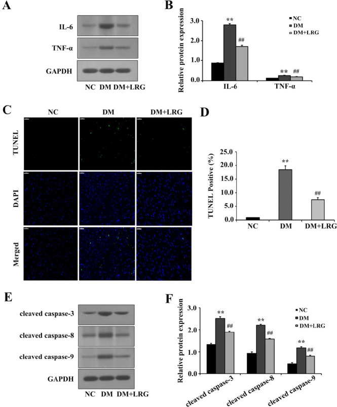
Figure 2.
Liraglutide reduces myocardial inflammation and pyroptosis in diabetic rats. (A) Representa-tive images of Western blotting; (B) Interleukin (IL)-6 and tumor necrosis factor (TNF)-α ex-pression; (C) Representative images of myocardium tissue sections stained with TUNEL (×400, bar=50 µm). The pyroptotic cells were detected via TUNEL staining (green), and the nuclei were detected using DAPI (blue); (D) Quantitative data for the TUNEL-positive my-ocytes; (E) Representative images of Western blotting; (F) Cleaved caspase-3, cleaved caspa-se-8, and cleaved caspase-9 expression. Data are presented as the mean±SEM, n=5–10 per group. **P<0.01 vs NC; ##P<0.01 vs DM
NC: normal control; DM: diabetes mellitus; LRG: liraglutide

