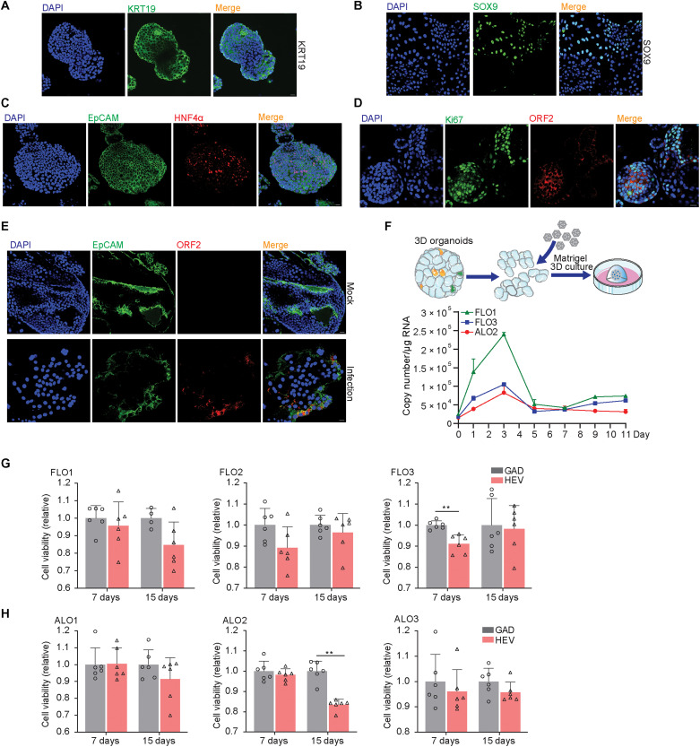
Fig. 3. Immunostaining of relevant markers, HEV infection in polarized organoid cells with cholangiocyte phenotype, and the effects of HEV replication on organoid growth.
(A) Immunofluorescence staining of KRT19 in ICOs. (B) Immunofluorescence staining of SOX9 in organoids. (C) Immunofluorescence staining of hepatic marker HNF4α in organoids. (D) Immunofluorescence staining of proliferation marker Ki67. (E) Immunofluorescence staining for viral ORF2 protein in ICOs on day 3 after inoculation of infectious HEV particles. (F) Virus growth curve by inoculation of organoids with HEV particles. Virus titers of 1 hour after inoculation was set as day 0 as starting point. (G and H) Cell viability of organoids electroporated with p6 RNA and HEV RNA with GAD mutation. Cell viability is tested on days 7 and 15 after electroporation (n = 4 to 6).

