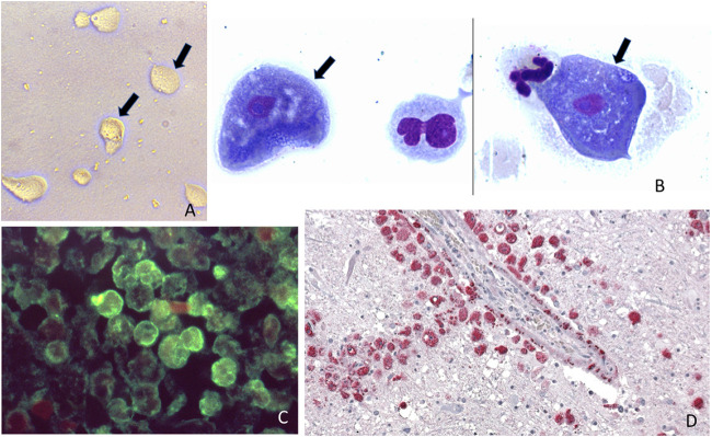
FIG 1.
Different diagnostic methods used for free living amoeba: (A) Free-living amoeba trophozoites observed in a culture using a non-nutrient agar and a bacterial lawn (arrows mark the trophozoites). (B) Giemsa stain of cerebrospinal fluid in a patient with Acanthamoeba spp. granulomatous amoebic encephalitis (arrows mark 2 trophozoites from the same slide but from different locations). (C) Immunofluorescence assay in brain tissue of a patient with N. fowleri (amoeba stained green). (D) Immunohistochemical assay in brain tissue of a patient with B.mandrillaris granulomatous amoebic encephalitis, red staining corresponds to the amoeba. Note that the amoeba surrounds a blood vessel. Panels A and C are from the Public Health Image Library, CDC.

