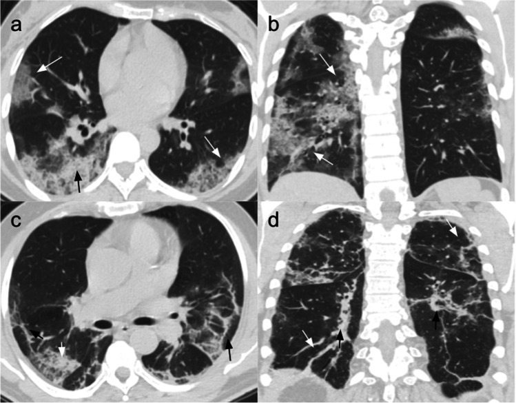
Fig. 1.
Common chest CT findings in COVID-19 pneumonia. Patient 1: CT scans of a 47-year-old woman affected by COVID-19 pneumonia and hospitalized for 6 days without ICU admission. She was treated with antiviral and antibiotic therapy, hydroxychloroquine, and low flow nasal cannula (2 ml/min). (a) Non-contrast CT scan, axial plane, performed at admission showing bilateral crazy-paving opacities (white arrows) and right posterior consolidation (black arrow). (b) Non-contrast coronal plane showing bilateral asymmetric GGOs and crazy-paving areas (white arrows), mostly in the posterior subpleural lung regions. Patient 2: CT scans of a 73-year-old man with COVID-19 pneumonia, hospitalized for 12 days without ICU admission. He was treated with a low flow nasal cannula (ranging from 2 to 4 ml/min), antibiotics, and IV fluids. (c) Non-contrast CT scan, axial plane, performed at admission showing bilateral GGOs with superimposed interlobular and intralobular septal thickening (white arrow), and architectural distortion appearing in the peripheral areas (black arrows). (d) Non-contrast coronal plane showing architectural distortion with bilateral subpleural lines (white arrows) and traction bronchiectasis (black arrows)

