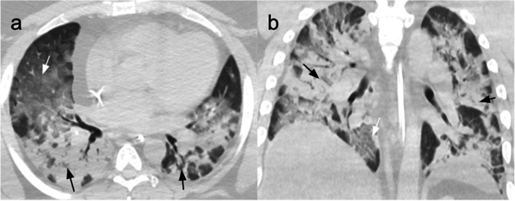
Fig. 2.
CT scans of a 36-year-old man affected by severe COVID-19 pneumonia and hospitalized for 11 days with ICU admission on the second day, after being treated with CPAP. In ICU, he went through seven cycles of pronation with progressive improvement of lung distress. (a) Non-contrast CT scan performed on the first day in ICU, axial plane, showing GGOs (white arrow) and consolidation (black arrows) in all the lobes, with only a few areas of normal parenchyma. (b) Non-contrast coronal plane CT scan showing diffuse bilateral consolidation crazy-paving pattern involving the majority of the lung parenchyma

