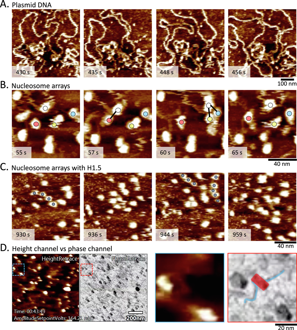
Figure 4.

Examples of chromatin sample imaged by HS-AFM. (a) Plasmids in fluid show dynamic behavior (Supplemental Video 1). (b) Tracking an individual nucleosome (white, red, green, and yellow circles), intermittent contact between neighboring nucleosomes were observed (Supplemental Video 2). (c) H3 chromatin compaction induced by H1 shows highly mobile nucleosomes that form temporary nucleosome phasing (dark blue circles; Supplemental Video 3). (d) The height and phase channels provide complementary data. In particular, the phase channel allows for distinguishing lighter DNA from darker nucleosomes (see inset, where DNA (blue) is distinct from nucleosome (red); Supplemental Video 4).
