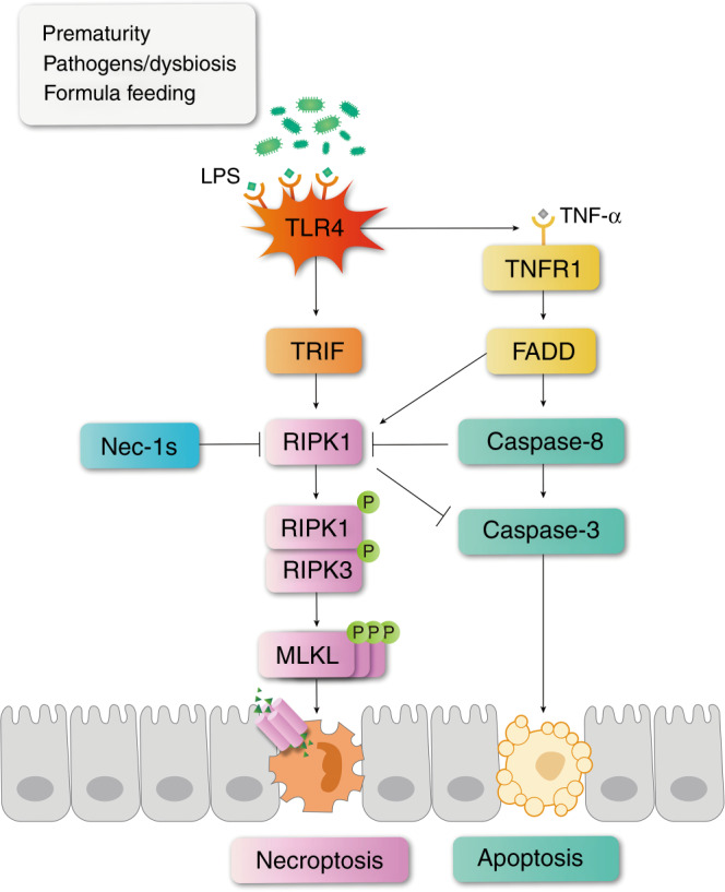
Fig. 8. Schematic illustrations of how necroptosis and apoptosis were regulated in NEC.

In NEC patients or cells challenged by LPS, TLR4 is activated. On the one hand, TLR4 binds to TRIF to activate necroptosis. On the other hand, TLR4 activation induces the production of inflammatory cytokines including TNF-α, which further binds with TNFR1 and leads to apoptosis and necroptosis simultaneously. When RIPK1 is inhibited by necrostatin-1s, or when TLR4 is removed, necroptosis becomes suppressed. The production of TNF-α is also reduced so that caspase-8 is decreased, leading to a decreasing trend of apoptosis. Meanwhile, the suppression of RIPK1 might relieve its inhibitory effect on caspase-3, leading to an increased trend of apoptosis. These two effects may counteract each other, but in general necrostatin-1 and TLR4 knockout showed an alleviating effect on NEC mice.
