Figure 1.
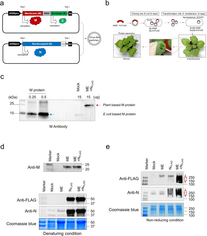
Expression of SARS-CoV-2 VLPs in N. benthamiana. (a) Schematic representation of two vectors for the formation of SARS-CoV-2 VLPs. pBYR2fp-MIRESE (upper) and pBYR2fp-NFLAG (bottom) vectors contained a recombinant gene, M-IRES-E (MIRESE) and N-FLAG (NFLAG). (b) Schematic representation of the co-expression system process by two vectors using the agroinfiltration method in tobacco plants. (c) Identification of M protein (~ 25 kDa) expression using Western blot analysis in total soluble protein (TSP) extracted from tobacco leaves 3 dpi of pBYR2fp-MIRESE. Recombinant M protein (15 kDa) derived from E. coli used as a positive control was loaded with 0.25 µg (line 2) and 0.5 µg (line 3), respectively. ME represents samples injected with pBYR2fp-MIRESE. Western blot analysis under both denaturating (d) and non-denaturating conditions (e) to confirm the co-expression of M, E, and N in TSP. NFLAG represents samples injected with pBYR2fp-NFLAG. ME + NFLAG represents samples co-injected with pBYR2fp-MIRESE and pBYR2fp- NFLAG. All images were cropped. See Supplementary Figs. S1–S3 for full size of blot.

