
Figure 7:
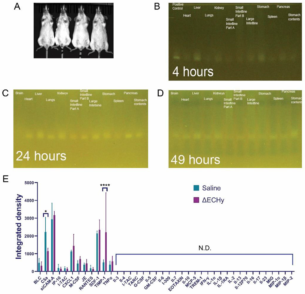
(A) Mice biodistribution study for ΔElux showing no detectable signals in-vivo (n=4). (B, C, D) PCR amplification for detecting the presence of ΔElux containing pakfplux1 plasmid from the microbiome genetic material isolated from each organ (121bp). (E) Graphical plot of cytokine profiling results from the biocompatibility study in mice. ΔECHy was compared with saline and only two cytokines out of the 40 tested were observed to be significantly regulated, complement component 5a (C5a, p=0.016) was downregulated and the tissue inhibitor of metalloproteinases (TIMP-1, p<0.0001) was upregulated. No other inflammatory markers were detected (n=3, data represented as mean±SD and compared using two-way anova (tukey’s multiple comparisons test).