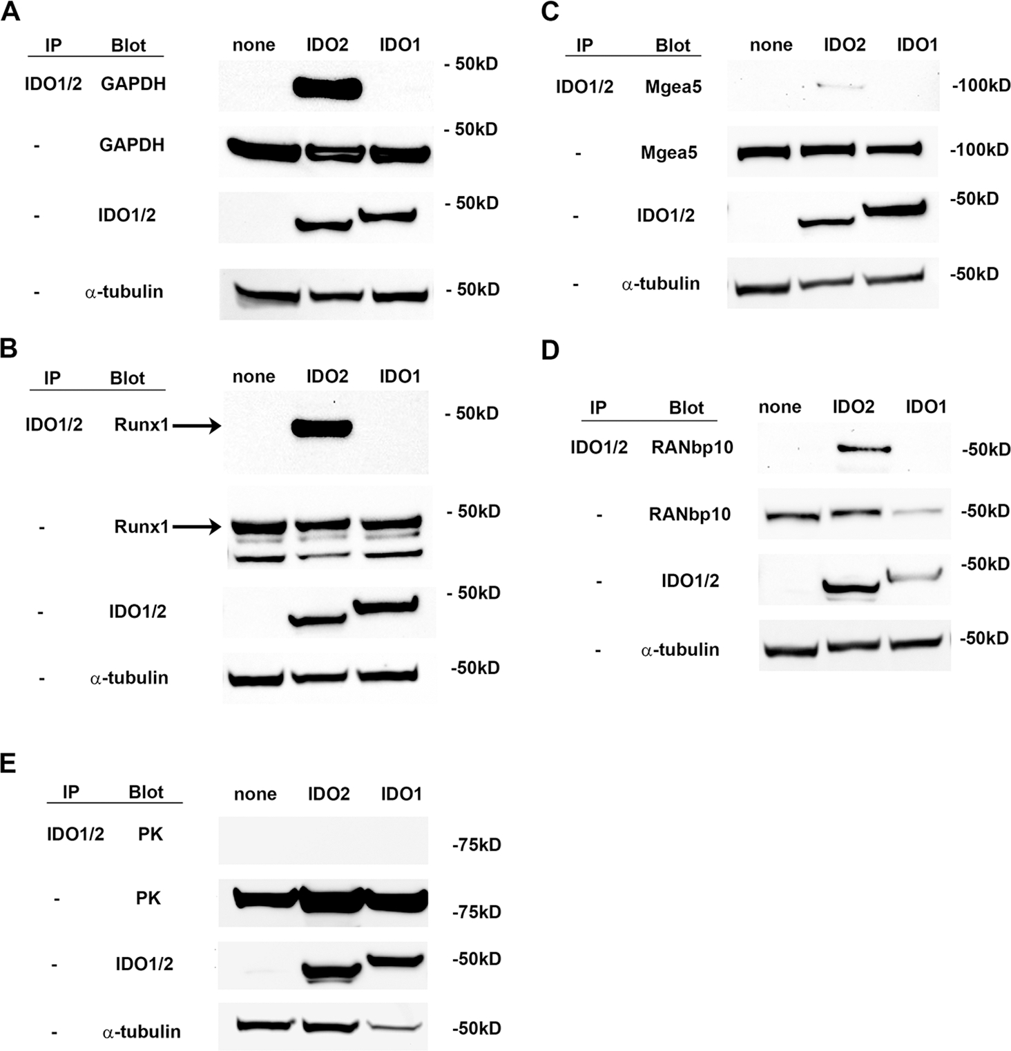
Figure 7. IDO2-binding protein interactions suggest potential non-enzymatic pathway mediated by IDO2.

293-T-REx™ cells, stably transfected with V5-tagged IDO2, IDO1 or no IDO (none), were transiently transfected with FLAG-tagged (A) GAPDH, (B) Runx1, (C) Mgea5, (D) RANbp10, or (E) PK. Lysates were immunoprecipitated with anti-V5 resin and then immunoblotted with anti-FLAG-HRP. Whole lysates were also directly blotted with anti-V5 or anti-FLAG to verify expression of the transfected proteins. Although the same plasmid backbone was used to express all four test proteins, there was variation in the expression level between the individual proteins (GAPDH > Runx1 and Mgea5 >RANbp10). However, each individual protein was expressed at approximately the same level between Trex cells expressing IDO2, IDO1, or no IDO. Representative blots are shown from a total of 2 independent experiments. Arrow indicates Runx1 band.
