Figure 1:

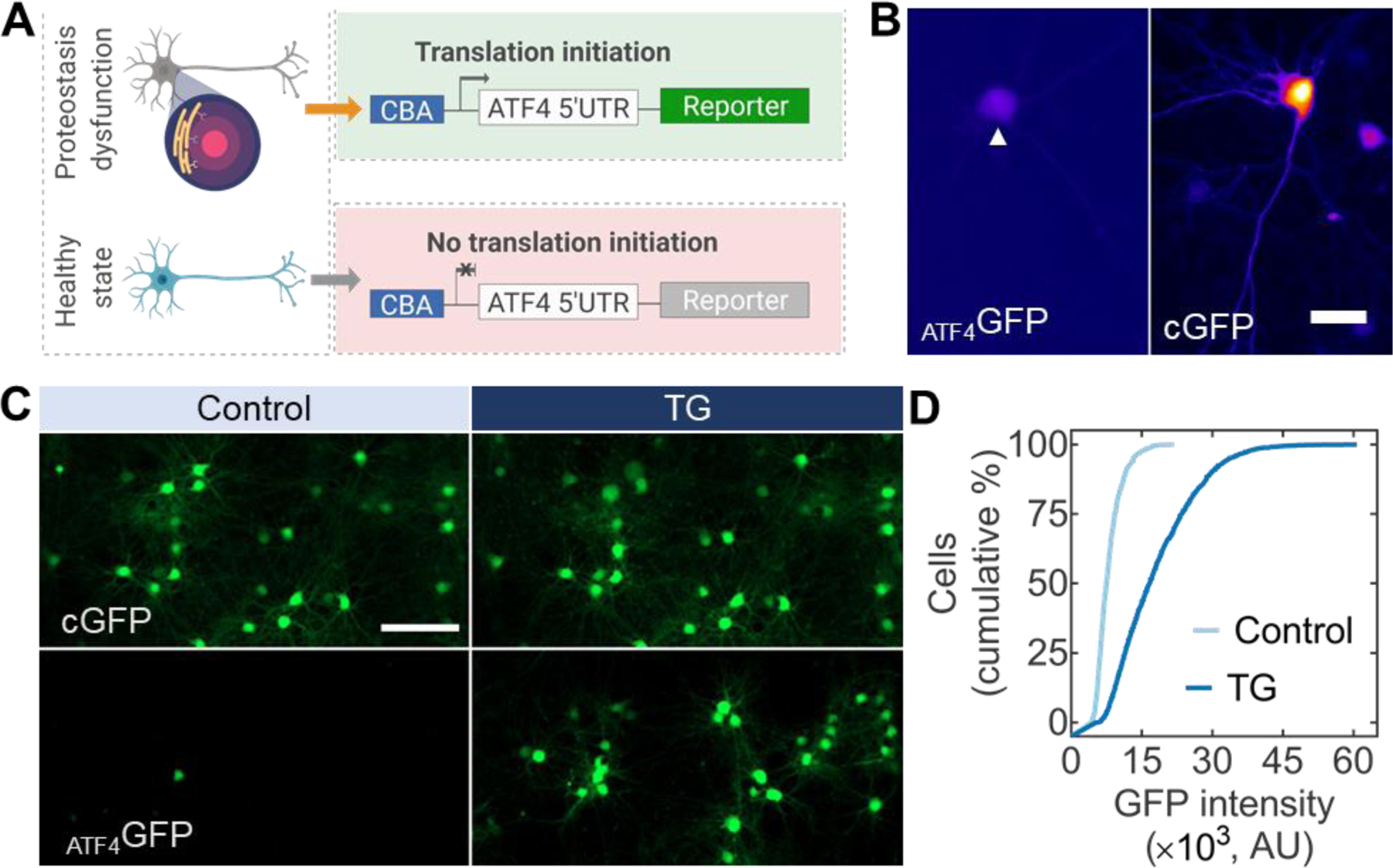
Stress-dependent transgene expression vectors applied in primary neurons. (A) AAV vectors with a fluorescent reporter downstream of a translational control operator from the ATF4 5’ UTR allow for the assessment of proteostasis dysfunction in neurons. (B) Primary cortical neurons under normal culture conditions seven days after viral transduction. Stress-dependent expression (ATF4GFP, left, arrow) is substantially lower than constitutively expressed GFP (CGFP, right). Scale bar 25 µm. (C) Primary neurons expressing CGFP (top) or ATF4GFP (bottom) untreated (left, control) or treated with TG for 24 h (right, TG). Scale bar 100 µm. (D) Population-based quantification of ATF4GFP induction from control following TG treatment. n > 3,000 neurons.
