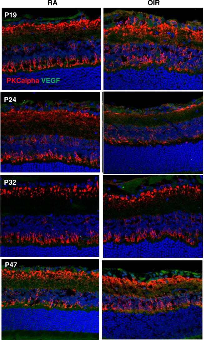
Fig. 2. VEGF-A165 expression and synaptic integrity in RA and OIR.

Vegfa165 antibody (green) staining was observed to be increased in both RA (n = 4) and OIR (n = 4) mice at P19. While some retinal areas in OIR mice appeared to have increased VEGF-A expression, there was no difference between total VEGF-A164 expression in RA and OIR mice at any age. PKCalpha (red) showed mis-aligned rod bipolar cells in OIR mice, while RA mice had uniformly aligned synapses. Vegfa165 vascular endothelial growth factor-a164, RA room air, OIR oxygen-induced retinopathy, P postnatal day age.
