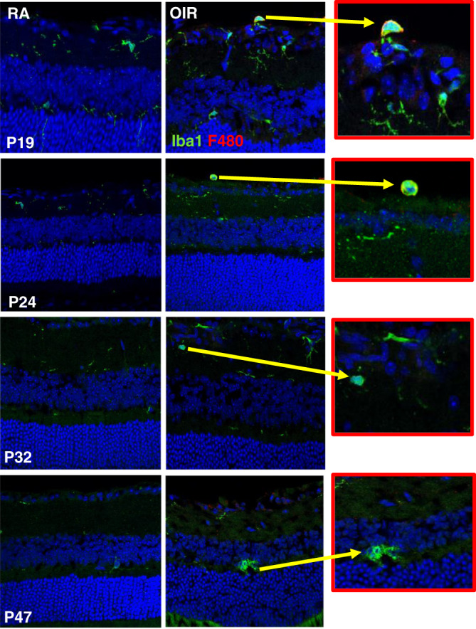
Fig. 3. Microglia activation in RA and OIR mice.

Two subsets of Iba1+ cells were noted in OIR mice: ramified, dendritic-appearing and amoeboid, round-appearing. Certain microglia subset co-stained both Iba1+ and F4/80+. Ameoboid, rounded F4/80+ microglia represented <5% of microglia. RA mice had sparse number of microglia, mainly in a ramified, dendritic phenotype, typical of resting microglia. In OIR mice, Iba1+ microglia was increased, highest at P19, and decreased with age. F4/80 staining was increased at P19 and P24 in OIR mice compared to RA mice, but similar at P32 and P47 in RA and OIR mice. Iba1-stained cells were higher in OIR compared to RA mice at every age tested. The ratio of F4/80 and Iba1 cells was increased at P24, with 30% higher proliferation of Iba1 cells. Yellow arrow shows a magnified view of activated amoeboid microglia in OIR mice that co-stained both Iba1+ and F4/80+. RA room air, OIR oxygen-induced retinopathy, P postnatal day age.
